From suspicion to surprise: a case of soft palate bulge diagnosed as parapharyngeal space pleomorphic adenoma
DOI:
https://doi.org/10.34631/sporl.3165Palavras-chave:
Parapharyngeal space tumors, pleomorphic adenoma, transoral excision, salivary gland tumor, soft palate swelling, benign neoplasmResumo
Introduction: Parapharyngeal space (PPS) tumors are rare and often present diagnostic challenges due to their deep anatomical location and nonspecific symptoms. Pleomorphic adenomas in this region are very rare, and when unrelated to the parotid gland, they can mimic malignancies.
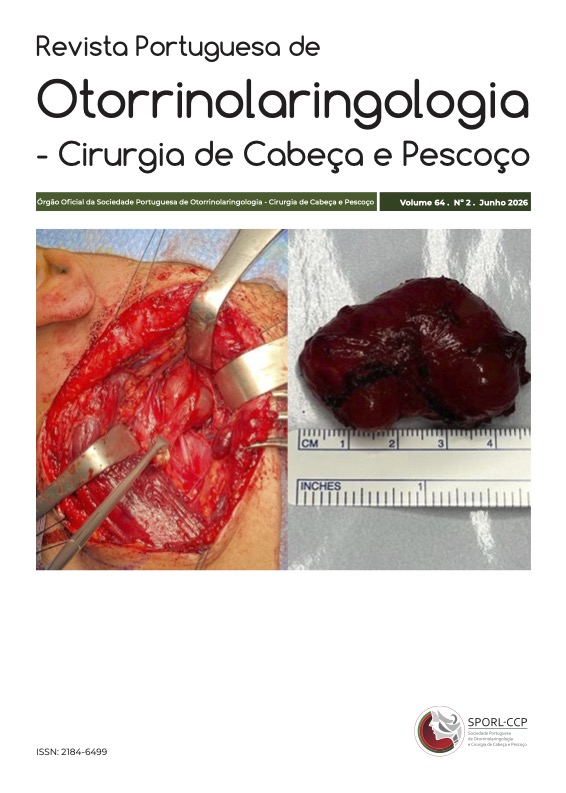
Case Presentation: A 51-year-old male presented with a 9-month history of progressive right-sided soft palate swelling, dysphagia, and hot potato voice. MRI revealed a solid-cystic mass in the prestyloid PPS, radiologically suspicious for malignancy and separate from the parotid gland. The lesion was excised via transoral endoscopic approach. Histopathology confirmed pleomorphic adenoma. Recovery was uneventful, with no recurrence on follow-up.
Conclusion: Pleomorphic adenomas of the PPS can mimic malignancy clinically and radiologically. Complete surgical excision via a transoral endoscopic approach can be safe and effective in selected patients. Histopathological confirmation is essential to guide management.
Downloads
Referências
1. Eveson JW, Auclair PL, Gnepp DR. Tumors of the salivary glands. In: Barnes L, Eveson JW, Reichart PA, Sidransky D, eds. World Health Organization Classification of Tumors: Pathology and Genetics of Tumors of the Head and Neck. Lyon: International Agency for Research on Cancer (IARC); 2005: 209-281.
2. Stambuk HE, Patel SG. Imaging of the parapharyngeal space. Otolaryngol Clin North Am. 2008 Feb;41(1):77-101, vi. doi: 10.1016/j.otc.2007.10.012.
3. Guntinas-Lichius O, Gabriel B, Klussmann JP. Risk of facial palsy and severe Frey's syndrome after conservative parotidectomy for benign disease: analysis of 610 operations. Acta Otolaryngol. 2006 Oct;126(10):1104-9. doi: 10.1080/00016480600672618.
4. Luna-Ortiz K, Navarrete-Alemán JE, Granados-García M, Herrera-Gómez A. Primary parapharyngeal space tumors in a Mexican cancer center. Otolaryngol Head Neck Surg. 2005 Apr;132(4):587-91. doi: 10.1016/j.otohns.2005.01.013.
5. Khafif A, Segev Y, Kaplan DM, Gil Z, Fliss DM. Surgical management of parapharyngeal space tumors: a 10-year review. Otolaryngol Head Neck Surg. 2005 Mar;132(3):401-6. doi: 10.1016/j.otohns.2004.09.062.
6. Rahnama M, Orzędała-Koszel U, Czupkałło L, Lobacz M. Pleomorphic adenoma of the palate: a case report and review of the literature. Contemp Oncol (Pozn). 2013;17(1):103-6. doi: 10.5114/wo.2013.33438.
Downloads
Publicado
Como Citar
Edição
Secção
Licença
Direitos de Autor (c) 2026 Deepalakshmi Tanthry, Ankitha M., V. Akarsh Hegde, M. Sapna, Anaghaa Bhat Nagri

Este trabalho encontra-se publicado com a Licença Internacional Creative Commons Atribuição-CompartilhaIgual 4.0.