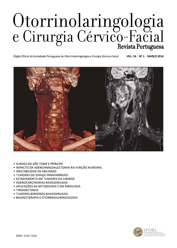
Parapharyngeal space tumors: A 10 year experience of IPO-LFG
DOI:
https://doi.org/10.34631/sporl.291Keywords:
Parapharyngeal space tumors, head and neck tumorsAbstract
Introduction: Parapharyngeal space neoplasms are rare, accounting for only 0.5% of head and neck tumors. The majority of PPS tumors are benign, but a wide spectrum of pathologies, both benign and malignant, has been encountered in this region. This range of histopathologies in combination with the complex anatomy of the parapharyngeal space creates complex diagnostic and management challenges.
Objective: To describe and analyze a case series of primary parapharyngeal space neoplasms at Instituto Português de Oncologia de Lisboa Francisco Gentil (IPOLFG).
Methods: Retrospective review of medical records of patients with parapharyngeal space neoplasms, diagnosed or referred to IPOLFG between 1st of January of 2003 and 31st of December of 2013. Results: 38 patients were included. The median age was 52 years (Interquartile range 40-63). 10 patients (26.3%) were asymptomatic. The most common symptom was an oropharyngeal lump sensation (23.7%). All patients had preoperative imaging: 94.7% computed tomography and 68.4% magnetic resonance image. 39.5% underwent fineneedle aspiration biopsy. 31 tumors were benign (81.6%), with pleomorphic adenomas comprising the majority (58.1%). 7 were malignant (18.4%), with carcinoma ex pleomorphic adenoma (28.6%) and lymphoma (28.6%) being the most common. 36 patients (94.7%) underwent primary surgical management; the other 2 patients (5,3%) were treated with chemotherapy and chemoradiotherapy, respectively. The cervical approach was the most common (80%). A mandibulotomy was required in just 5.7% of primary cases. The most frequent complication was cranial neuropathy, identified in 22,2%. Of these, 75% were sequelae from resection of neurogenic tumors. Median follow-up was 6.5 years.
Conclusion: PPS tumors require a high index of suspicion to diagnose them at an early stage. Complete surgical resection is the mainstay of treatment. The optimum surgical approach needs to be selected on an individual basis, but the cervical approach is safe and effective for most PPS neoplasms.
Downloads
References
Olsen KD. Tumors and Surgery of the Parapharyngeal space. Laryngoscope 1994; 104:1- 25.
Jones A. Tumors of the parapharyngeal space. Em: Gleeson M. Scott Brown´s otorhinolaryngology, head and neck surgery, 7th ed. Edward Arnold; 2008: 2522-39.
Bradley PJ, Bradley PT, Olsen KD. Update on the management of the parapharyngeal tumors. Curr Opin Otolaryngol Head Neck Surg 2011; 19: 92-98.
Gil Z, Fliss DM. Surgical management of parapharyngeal space tumors. Em: Souza C. Atlas of head and neck surgery, 1st ed. Jaypee; 2013: 319-31.
Starek I, Mihal V, Novak Z. Paediatric tumours of the parapharyngeal space. Int J Pediatr Otorhinolaryngol 2004; 68: 601–606.
Riffat F, Dwivedi R, Palme C, Fish B et al. A systematic review of 1143 parapharyngeal space tumors reported over 20 years. Oral Oncology 2014; 50: 421-30.
Cassoni A, Terenzi V, Della Monaca M, et al. Parapharyngeal space benign tumours: our experience. J Craniomaxillofac Surg. 2014; 42(2): 101-5.
Caldarelli C, Bucolo S, Spisni R, Destito D. Primary parapharyngeal tumors: a review of 21 cases. Oral Maxillofac Surg. 2014; 18(3): 283-92.
Kuet M, Kasbekar A, Masterson L, Jani P. Management of tumors arising from the parapharyngeal space: a systematic review of 1293 cases reported over 25 years. Laryngoscope. 2014 Dec 2. doi: 10.1002/lary.25077.
Hughes K, Olsen K, McCaffrey T. Parapharyngeal Space Neoplasms. Head and Neck 1995;17:124-130.
Som P, Biller H, Lawson W, et al. Parapharyngeal space masses: an updated protocol based upon 104 cases. Radiology. 1984; 153: 149-56.
Eisele D, Richmon J. Contemporary evaluation and management of parapharyngeal space neoplasms. The journal of laryngology and otology. 2013; 127: 550-55.
Douville N, Bradford C. Comparison of ultrasound-guided core biopsy versus fine-needle aspiration biopsy in the evaluation of salivary gland lesions. Head Neck, 2013; 35(11): 1657-61.


