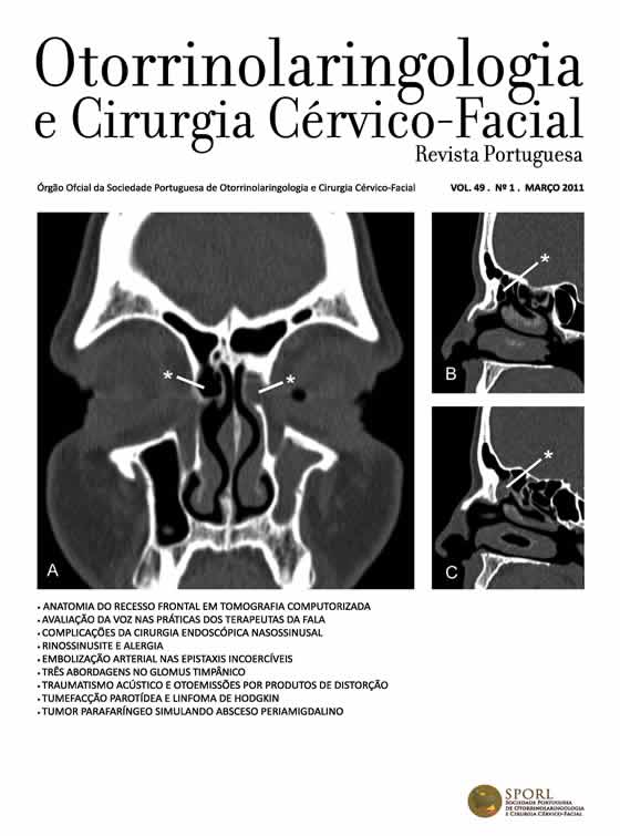
Computed tomography analysis of frontal recess anatomy: Study of 50 patients
DOI:
https://doi.org/10.34631/sporl.146Keywords:
frontal recess, accessory cells, anatomy, imagiologyAbstract
Objective: Study the prevalence of frontal recess accessory cells in patients with indication for endonasal surgery.
Study Design: Anatomical study of sinus CT scans.
Material and Methods: Analysis of sinus CT scans from patients with indications for endonasal surgery. Exclusion criteria included previous surgery, complicated sinusitis and disease that unable structures identification.
Results: 50 patients were included in the study with CT scans performed at diferent radiology centers. The prevalence of each cell was: Agger Nasi (92%), Frontoethmoidal cells type I (28%), type II (15%), type III (10%), Frontal Bulla (5,3%), Suprabullar cell (44,7%), Supraorbital cell(20%), Interfrontal septal cell (38%).
Conclusions: The study describes the frontal recess pneumatization pattern in patients with nasosinusal symptoms. The results were similar with those found in other studies.
Downloads
References
Arslan H, Aydınlıoğlu A, Bozkurt M, Egeli E. Anatomic Variations of the Paranasal Sinuses: CT examination for Endoscopic Sinus Surgery. Auris Nasus Larynx 1999; 26:39-48.
Stammberger H. Functional Endoscopic Sinus Surgery: The Messerklinger Technique. Philadelphia: BC Decker, 1991:60-87.
Friedman M, Bliznikas D, Vidyasagar R, Landsberg R. Frontal Sinus Surgery 2004: Update of Clinical Anatomy and Surgical Techniques. Operative Techniques Otolaryngol Head Neck Surg 2004 Mar;15(1):23-31.
Landsberg R, Friedman M. A Computer-Assisted Anatomical Study of the Nasofrontal Region. Laryngoscope 2001 Dec;111:2125-30.
Lee W, Kuhn F, Citardi M. 3D Computed Tomographic Analysis of Frontal Recess Anatomy in patients without frontal sinusitis. Otolaryngol Head Neck Surg 2004 Sep;131(3):164-73.
Otto K, DelGaudio J. Operative findings in the frontal recess at time revision surgery. Am J Otolaryngol 2010;31:175-80.
Cho J, Citardi M, Lee W, Sautter N et al.. Comparison of frontal pneumatization patterns between Koreans and Caucasians. Otolaryngol Head Neck Surg 2006 Nov;135(5):780-86.
Beale T, Madani G, Morley S. Imaging of the Paranasal Sinuses and Nasal Cavity: Normal Anatomy and Clinically Relevant Anatomical Variants. Semin Ultrasound CT MR 2009;30:2-16.
Kantarci M, Karasen R, Alper F, Onbas O et al.. Remarkable anatomic variations in paranasal sinus region and their clinical importance. Eur J Radiol 2004;50:296-302.
Keast A, Yelavich S, Dawes P, Lyons B. Anatomical variations of the paranasal sinuses in Polynesian and New Zeland European computerized tomography scans. Otolaryngol Head Neck Surg 2008 Aug;139(2):216-21.
Wormald PJ. Three-dimensional building block approach to understanding the anatomy of the frontal recess and frontal sinus. Operative Techniques Otolaryngol Head Neck Surg 2006 Mar;17(1):2-5.
Ercan İ, Çakır B, Sayın İ, Başak M et al.. Relationship between the superior attachment typi of uncinate process and presence of agger nasi cell: A computer-assisted anatomic study. Otolaryngol Head Neck Surg 2006; 134(6): 1010-14.
Jacobs JB, Lebowitz RA, Sorin A, et al.. Preoperative sagital CT evaluation of the frontal recess Am J Rhinol 2000;1:33-7.
Wormald PJ. The agger nasi cell: the key to understand the anatomy of the frontal recess. Otolaryngol Head Neck Surg 2003;129:497-507.
Van Alyea OE. Ethmoid labyrinth: anatomic study with consideration of the clinical significance of its structural characteristics. Arch Otolaryngol Head Neck Surg 1939;29:881-901.
Bolger WE, Butzin CA, Parson DS. Paranasal sinus body anatomic variations and mucosal abnormalities: CT analysis for endoscopic sinus surgery. Laryngoscope 1991; 101:56-64.
Owen R, Kuhn F. Supraorbital ethmoid cell. Otolaryngol Head Neck Surg 1997;116:254-61.
Zhang L, Han D, Ge W, Tao J et al.. Computed tomographic and endoscopic analysis of supraorbital ethmoid cells. Otolaryngol Head Neck Surg 2007;137:562-68.
Alquezar MP, Liesa RF, Muñoz AL, Delgado LP et al.. La arteria etmoidal anterior en el laberinto etmoidal: Revisión bibliográfica sobre variantes anatómicas y referencias para la cirurgia endoscópica. Acta Otorrinolaringol Esp 2010 (in press).