Sucesso cirúrgico da reparação de fístulas de LCR da base anterior do crânio por via endoscópica
DOI:
https://doi.org/10.34631/sporl.2185Palavras-chave:
Fístula de LCR, underlay, overlay, retalho nasosseptal, retalho livre de mucosaResumo
Introdução: As fístulas de líquido cefalorraquidiano (LCR) são comunicações anormais entre o espaço subaracnóideu e a cavidade nasal e podem ser classificadas em espontâneas e traumáticas (incluindo as fístulas iatrogénicas). Várias técnicas e materiais têm sido descritos na reparação endoscópica de fístulas de LCR. No entanto, a técnica endoscópica de eleição continua a ser um tema amplamente debatido.
Objetivo: Apresentar a experiência de um centro terciário na reparação fístulas de LCR da base anterior do crânio.
Métodos: Estudo retrospetivo realizado num hospital terciário, entre janeiro de 2012 e dezembro de 2023.
Foram incluídos todos os doentes submetidos a reparação endoscópica de fístulas de LCR da base anterior do crânio. Foram recolhidos dados demográficos, sintomas, exames de diagnósticos e fatores intraoperatórios. Foi realizada análise estatística descritiva e analítica.
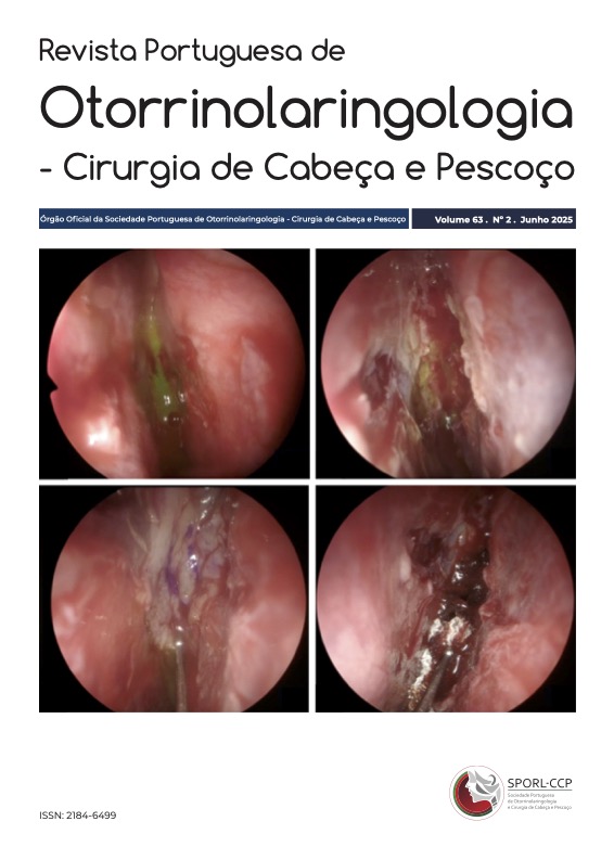
Resultados: Vinte e nove doentes (19 mulheres, idade média de 59,6±13 anos) com 30 fístulas de LCR foram incluídos. A etiologia mais comum foi a espontânea, responsável por 59% de todas as fístulas de LCR, seguida da iatrogénica (31%) e da traumática (10%). Sete doentes com fístula de LCR espontânea eram obesos, sendo que um deles apresentava estigmas de hipertensão intracraniana idiopática. As fístulas de LCR espontâneas foram mais frequentemente localizadas na lâmina crivosa e na lamela lateral. A lamela lateral foi o local mais frequentemente envolvido nas fístulas de LCR iatrogénicas. O tamanho médio do defeito foi de 5,8±7,4 mm. A drenagem lombar foi colocada em 12 doentes antes da cirurgia. Utilizamos a técnica under-overlay em 63% dos casos, overlay em 33% e em 3% (um caso) utilizamos uma abordagem combinada (endocraniana e endonasal). A técnica foi escolhida de acordo com a etiologia e tamanho do defeito, e preferência do cirurgião. Na técnica under-overlay, os materiais mais utilizados foram a duragen® underlay e retalhos livres de mucosa overlay selados com cola de fibrina.
Quanto à técnica overlay, utilizamos retalhos livres de mucosa na maioria dos casos, Surgicel® e cola de fibrina. A média do número de camadas por doente foi de 3,9±1,2.
As fístulas de LCR da lâmina crivosa foram frequentemente encerradas por técnica exclusivamente overlay, pela incapacidade de disseção dural sem aumentar significativamente o tamanho do defeito ósseo. Não ocorreram complicações pós-operatórias. A taxa de sucesso foi de 90%. Houve necessidade de re-intervenção em apenas um caso.
Conclusão: A reparação de fístulas de LCR por via endoscópica é uma técnica segura e eficaz. A alta taxa de sucesso observada no nosso estudo revela que a técnica cirúrgica utilizada deve ser decidida de acordo com a etiologia, tamanho e localização do defeito na base do crânio, sendo que nenhuma técnica se mostrou significativamente superior.
Downloads
Referências
Majhi S, Sharma A. Outcome of endoscopic cerebrospinal Ffuid rhinorrhoea repair: an institutional study. Indian J Otolaryngol Head Neck Surg. 2019 Mar;71(1):76-80. doi: 10.1007/s12070-018-1485-2.
Murray RD, Friedlander R, Hanz S, Singh H, Anand VK, Schwartz TH. Nonrandom spatial clustering of spontaneous anterior fossa cerebrospinal fluid fistulas and predilection for the posterior cribriform plate. J Neurosurg. 2017 May;126(5):1720-1724. doi: 10.3171/2016.4.JNS152975
Fiore G, Bertani GA, Carrabba GG, Guastella C, Marfia G, Tariciotti L. et al. “The ‘parachute’ technique for the endoscopic repair of high-flow anterior skull-base CSF leaks. World Neurosurg. 2021 Jul:151:e880-e887. doi: 10.1016/j.wneu.2021.05.006
Sigler AC, D'Anza B, Lobo BC, Woodard TD, Recinos PF, Sindwani R. Endoscopic skull base reconstruction: an evolution of materials and methods. Otolaryngol Clin North Am. 2017 Jun;50(3):643-653. doi: 10.1016/j.otc.2017.01.015.
Kim-Orden N, Shen J, Or M, Hur K, Zada G, Wrobel B. Endoscopic endonasal repair of spontaneous cerebrospinal fluid leaks using multilayer composite graft and vascularized pedicled nasoseptal flap technique. Allergy Rhinol (Providence). 2019 Nov 13:10:2152656719888622. doi: 10.1177/2152656719888622.
Kim BK, Kong DS, Nam DH, Hong SD. Comparison of graft materials in multilayer reconstruction with nasoseptal flap for high-flow CSF leak during endoscopic skull base surgery. J Clin Med. 2022 Nov 13;11(22):6711. doi: 10.3390/jcm11226711.
Khatiwala RV, Shastri KS, Peris-Celda M, Kenning T, Pinheiro-Neto CD. Endoscopic endonasal reconstruction of high-flow cerebrospinal fluid leak with fascia lata "button" graft and nasoseptal fflap: surgical technique and case series. J Neurol Surg B Skull Base. 2020 Dec;81(6):645-650. doi: 10.1055/s-0039-1693124
Hoerter JE, Kshirsagar RS. Nasoseptal Flap. [Updated 2023 Jul 12]. In: StatPearls [Internet]. Treasure Island (FL): StatPearls Publishing; 2024 Jan-. Available from: https://www.ncbi.nlm.nih.gov/books/NBK576383/
Downloads
Publicado
Como Citar
Edição
Secção
Licença
Direitos de Autor (c) 2025 Joana Guincho, Luís Baptista, Filipe Correia, Rui Cabral, Pedro Escada

Este trabalho encontra-se publicado com a Licença Internacional Creative Commons Atribuição-CompartilhaIgual 4.0.